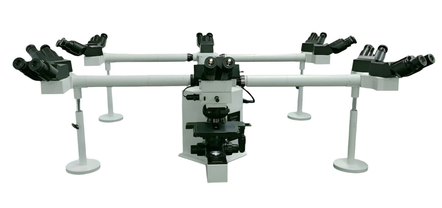
The R-series multi-view biological microscope is a teaching-oriented microscopic observation system developed by Abner Micro-Nano Technology for medical teaching, biological experiment teaching and professional training scenarios. Built on a high-performance infinity-corrected optical system, this series adopts a synchronous multi-observation head co-view structure design, enabling multiple observers to view the same microscopic field of view simultaneously and equivalently, which effectively guarantees the consistency of teaching demonstrations, experiment interpretation and results.
The R-series microscope is particularly suitable for application scenarios with high requirements for "synchronous observation" and "unified field of view", such as classroom teaching, skill training, pathology and sample interpretation. It can significantly improve teaching efficiency and demonstration quality.
### I. Product Features
Multi-person synchronous viewing design
Supports 2–10 users (customizable) to observe the same field of view simultaneously, ensuring consistent teaching and interpretation results.
Infinity-corrected optical system
Delivers sharp imaging with well-controlled aberrations, guaranteeing uniform image quality across multiple observation heads.
Modular observation head assembly
Flexible combination of a main observation head and multiple auxiliary observation heads, supporting left-right / front-back layouts to adapt to different teaching spaces.
Ergonomic design for teaching efficiency
Optimized viewing angle to reduce fatigue during extended teaching and observation sessions.
Digital teaching expansion capability
Optional digital camera and display system to enable combined teaching via direct visual observation and screen display.
II. Technical Parameters (Typical Configuration)
Optical System
Optical System: Infinity-corrected optical system; Observation Mode: Upright biological microscope observation; Imaging Mode: Brightfield / Digital Imaging (Optional)
Multi-viewing System
Viewing Method: Beam-splitting multi-observation head structure; Standard Number of Users: 2–5 persons; Maximum Supported Users: ≤ 10 persons (Customized); Field of View Consistency: Uniform image brightness and clarity at all observation positions; Beam Splitting Ratio: Optimized beam splitting design to prioritize brightness for the main observation position
Eyepiece System
Eyepiece Type: High-eye-point widefield eyepiece; Standard Magnification: WF10×; Field Number: ≥ 18 mm
Objective Lens System
Objective Type: Plan Achromatic Objective Lenses
Standard Magnification Configuration:
4× / 0.10; 10× / 0.25; 40× / 0.65 (Spring); 100× / 1.25 (Oil, Spring, Optional); Objective Interface: Standard RMS Interface
Objective Revolver
Type: Inclined objective revolver; Number of Positions: 4-position / 5-position (Optional)
Stage
Type: Double-layer mechanical moving stage; Stage Size: Approx. 160 × 140 mm; X/Y Travel Range: ≥ 75 × 50 mm
Focusing System
Focusing Method: Coaxial coarse / fine focusing; Fine Focusing Precision: ≤ 2 μm; Anti-collision Protection: Built-in limit mechanism
Illumination System
Illumination Method: Transmitted illumination
Light Source Type: High-brightness LED cold light source
Brightness Adjustment: Continuously adjustable
Light Source Lifespan: ≥ 20,000 hours
Digital Teaching (Optional)
Camera Interface: C-mount
Supported Devices: USB / HDMI microscope cameras
Display Method: Monitor / Projection System
Software Functions: Real-time display, image capture, video recording, basic measurement
Electrical and Environmental Conditions
Power Supply: AC 100–240 V, 50/60 Hz
Operating Temperature: 5–40 ℃
Relative Humidity: ≤ 80% (non-condensing)
III. Typical Application Scenarios
Medical and biological experiment teaching and demonstration; joint interpretation of pathological samples; training for hospitals and inspection institutions; skill assessment and teaching demonstration; specimen analysis and discussion-based teaching
Display Images


