
CSL Laser Confocal Scanning Microscope
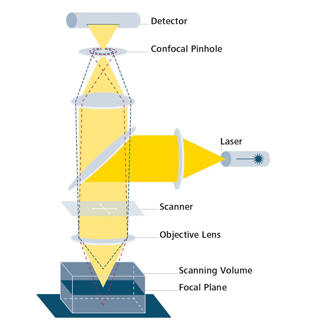
The CSL Laser Confocal Scanning Microscope is a high-performance confocal imaging system developed by Aibona Micro-Nano Technology for life science research, cellular imaging, 3D tissue reconstruction and functional microscopic imaging experiments. Based on the principles of point-by-point laser excitation and confocal pinhole imaging, the system adopts high-speed galvanometer scanning paired with high-sensitivity detection to capture high-contrast, high-resolution optical sectional images, and supports 3D volume reconstruction and multi-channel fluorescence imaging. Equipped with advanced laser light sources, precision optical scanning systems and an intelligent control software platform, the CSL series is widely applied in research fields including cell biology, developmental biology, histopathology, neural imaging and drug screening.
I. Core Advantages
High-Definition Confocal Section Imaging
Point-by-point laser scanning and confocal pinhole technology block out-of-focus light, greatly improving image contrast and depth discrimination.
Multi-Channel & Multi-Wavelength Fluorescence Imaging
Supports combined excitation wavelengths across 2 to 6 channels to achieve multi-label imaging of complex specimens.
3D Tomography & 3D Reconstruction
Rapidly acquires Z-stack layered data to reconstruct 3D sample structures and perform volumetric analysis.
High-Speed Scanning & Low Phototoxicity Imaging
High-speed galvanometers and advanced acquisition algorithms reduce light exposure, ideal for capturing dynamic processes of living cells.
Precision Control & Data Processing Software
The bundled scientific-grade software features real-time preview, image processing, 3D reconstruction, quantitative analysis, batch processing and data management functions.
II. Technical Specifications (Typical Configuration)
1) Laser Light Source System
Laser Type: Solid-state laser / Multi-line laser combinationTypical Excitation Wavelengths:405 nm, 488 nm, 561 nm, 638 nmOutput Stability: ±1% (typical)Optical Path Adjustment: Automatic / Manual (optional)
2) Scanning & Imaging System
Scanning Mode: Dual galvanometer high-speed scanning systemMinimum scanning pixel pitch: Determined by objective NA and wavelengthMaximum pixel acquisition rate: ≥ 4 megapixels (depends on camera/detector configuration)Average frame rate: Up to 30 fps (depends on resolution and channel count)
3) Detector
Detection Unit: Photomultiplier Tube (PMT) / High-sensitivity GaAsP detectorSensitivity: High quantum efficiencyNumber of Channels: 2–6 expandable channelsExcellent noise resistance, suitable for low-signal and low-light imaging
4) Optical System
Objective Interface: Standard infinity-corrected objective interfaceCommon Objective Magnifications: 10× / 20× / 40× / 60× (oil / water / dry)Typical Numerical Aperture (NA): 0.45–1.4 (varies by objective model)Adopts telecentric optical design to reduce optical distortion
5) Z-Axis Control
Z Step Drive: Motorized micro-step drive / Piezoelectric drive (optional)Minimum Z Step Size: ≤ 0.1 µm (piezoelectric version)Z Travel Range: Up to 20 mm (subject to sample chamber configuration)
6) Image Acquisition & Processing
Control Software: Research-grade data acquisition and image analysis softwareReal-time Display: Multi-channel overlay visualizationImage Processing: Denoising, gamma correction, contrast adjustment3D Reconstruction: Volume rendering, isosurface rendering, slice browsingBatch Processing: Automated Z-stack acquisition, time-lapse series capture
7. Expansion & Peripheral Accessories
Temperature control / Incubator (optional): For live cell imagingMotorized filter wheel: Automatic multi-channel switchingOptical path stabilizer: Automatic light intensity compensation
8) Electrical & Environmental Requirements
Power Supply: AC 100–240 V, 50/60 HzOperating Temperature: 18–28 ℃ (constant temperature & humidity environment recommended)Relative Humidity: 30%–70% (non-condensing)
III. Typical Application Fields
Imaging of cellular structures and processes; multiplex fluorescent labeling analysis; three-dimensional tomography and structural reconstruction of tissues; tracking of dynamic processes in living cells; research on developmental biology and neural network structures; mechanisms of drug action and high-content screening.


